Ten Parts Of Skull Medical Teaching Model
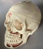
The skull model with detachable parts is an excellent educational tool for teaching anatomy to students in the field of dentistry and medicine.
The model is made of a resin material, which ensures its durability and longevity. The duplicates type II~III hardness resin material used to create the skull model is close to the human skeleton, making it a realistic representation of the human skull.
Feature
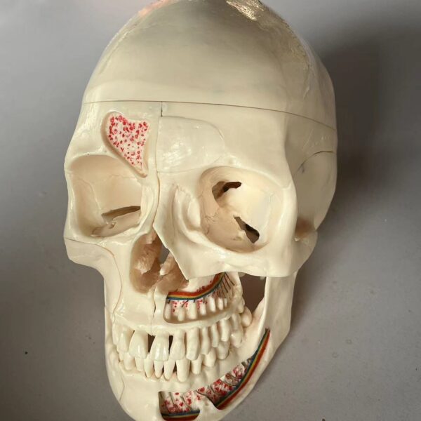
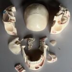
This model features ten detachable parts, allowing students to study each component of the skull individually.
The frontal sinus, perpendicular lamina, and vomer are equipped with flaps that can be opened to view the lateral nose wall and sphenoidal sinus.
On the left side, the temporal bone can be removed and folded up to expose the area of the tympanic membrane.
The maxilla and mandible of the skull are opened to reveal the alveolar nerves.
On the right side of the model, the temporal bone is opened to reveal the sigmoid sinus, the facial nerve canal, and the semicircular ducts.
These features make the model a comprehensive representation of the human skull’s anatomy, allowing students to study the intricate details of the skull’s structure.
Benefit in medical and dentistry teaching
- Comprehensive Understanding of the Anatomy: The skull model provides a detailed understanding of the human skull’s anatomy, including the bones, muscles, nerves, and blood vessels. This knowledge is essential for dental students, as it helps them understand the complex structures involved in oral health and dental procedures.
- Hands-on Learning: The detachable parts of the skull model allow students to learn through hands-on experience, which enhances their understanding of the subject. They can explore the structure of the skull by manipulating its parts, giving them a more immersive and engaging learning experience.
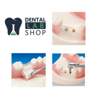
- Improved Clinical Skills: The Skull Model is an excellent tool for developing clinical skills. By studying the model, students can gain a better understanding of the different techniques used in dental procedures such as extractions, implants, and root canal treatments.
- Enhanced Patient Care: A thorough understanding of the skull’s anatomy is crucial for providing effective dental care to patients. By using the skull model in their education, dental students can develop a better understanding of the structures involved in oral health and how they interrelate. This knowledge can help them provide better patient care and improve treatment outcomes.
- Better Diagnosis and Treatment Planning: The skull model is an invaluable tool for diagnosis and treatment planning. By studying the model, students can develop a better understanding of the relationship between the different structures of the skull and how they affect oral health. This knowledge can help them diagnose and treat dental conditions more effectively.

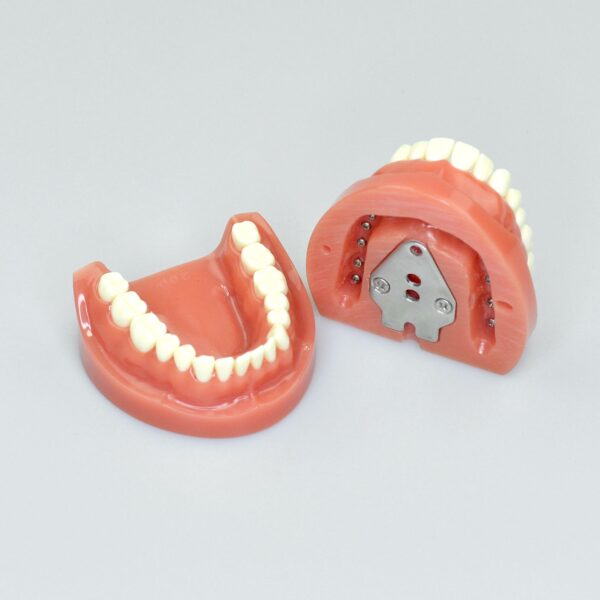


 Mode base has magnet plate, easy to attache simulator.
Free shipping.
Mode base has magnet plate, easy to attache simulator.
Free shipping.