

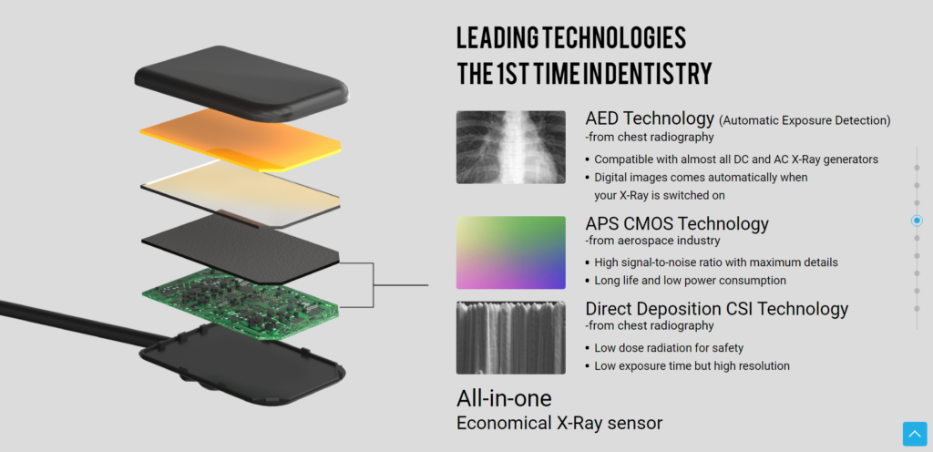
Digital x-ray sensors are an advanced imaging technology used in dentistry to capture digital images of teeth and gums. They detect radiation given off by an x-ray machine and convert it into electrical signals which are then sent to a computer. This allows for higher quality images with less radiation exposure, making them safer for patients.
The sensors are more efficient than traditional film-based x-rays and come in two sizes. Furthermore, multiple re-use and cost savings make digital x-ray sensors a reliable and durable choice for dental imaging.
When a dental X-ray sensor is used, a readable image is produced.
X-ray sensors can be used to detect decay, identify impacted teeth, examine tooth position, detect periodontal disease, and diagnose other dental conditions. X-ray sensors can be used to help diagnose and treat a variety of conditions, such as cavities, gum disease, abscesses, and jawbone issues. They can also be used to plan orthodontic treatments or dental implants.
Digital x-ray sensors provide many advantages over x-ray film for dental image captures.
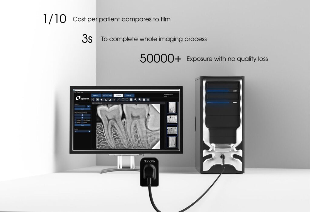
They allow faster image acquisition and display by software, image enhancement capabilities, lower radiation exposure, instant image storage, sharing and analysis, no chemical processing costs or hazards, multiple re-use and better diagnostic accuracy. Digital sensors also save time and can be purchased in optional sizes.
Most dental suppliers provide two size options for digital X-ray size:
– 4.4mm Thickness & 20x30mm Effective Imaging Area (for children)
– 4.4mm Thickness & 26x36mm Effective Imaging Area (for adults).
Multiple usage, cost saving
The digital x-ray sensor is a cost-saving alternative to x-ray film, and its usage data proves its reliability and durability: it has undergone 50,000 exposures without any loss of quality, and 70,000 successful bending tests.
The installed software process the images in a simple manner:
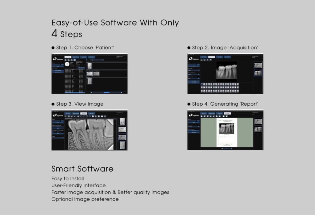
1. Place the sensor in the patient’s mouth and ensure that it is comfortable and secure.
2. Take the necessary radiographs using the appropriate exposure settings.
3. Ensure that the image is clear and of good quality.
4. Remove the sensor and store it properly.
5. Review the images and make any necessary adjustments.
6. Discuss the results with the patient and answer any questions they may have.
7. Store the images securely.
Technical table:
| Parameter | Small Sensor | Large Sensor |
|---|---|---|
| Sensor Dimensions (mm) | 25.4×36.8×4.4 | 30.4×41.9×4.4 |
| Effective Imaging Area (mm2) | 20×30 | 26×36 |
| Thickness | 4.4mm | 4.4mm |
| Imaging Technology | APS CMOS | APS CMOS |
| Image Processing Time | 3 seconds | 3 seconds |
| Bending Reliability Tests Passed | 70,000+ | 70,000+ |
| Exposure Detection | Automatic Exposure Detection | Automatic Exposure Detection |
| Signal-to-Noise Ratio | High | High |
| Sensor Lifespan | Long | Long |
| Power Consumption | Low | Low |
| Radiation Type | Low Radiation | Low Radiation |
| Resolution | 20 lp/mm | 20 lp/mm |
| Analog-to-Digital Conversion (AD) | 16-bit | 16-bit |
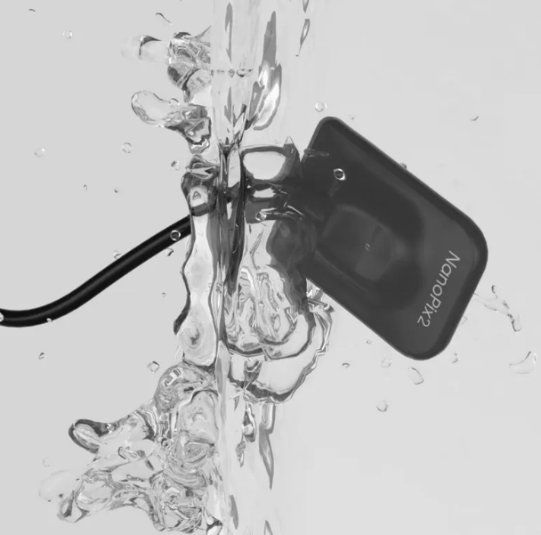
| Water and Dust Protection Rating | IP68 (Highest) | IP68 (Highest) |
| Data Connection | USB 2.0 | USB 2.0 |
| Manufacturer’s Warranty | 5 years (as applicable) | 5 years (as applicable) |
| Pre-installed Software | Included on USB Drive | Included on USB Drive |
| Cable Length | 3 meters | 3 meters |
| Manual | 1pcs | 1pcs |
| X-Ray Sensor | 1pcs | 1pcs |
| Software pre-installed in U-Key storage | 1pcs | 1pcs |
Conclusion
The ability to instantly capture, view, and enhance images leads to faster diagnoses and treatment planning.
Reduced radiation exposure also makes digital x-rays a safer choice for patients.
Cost savings from the lack of film and chemical processing along with the reuse capability of sensors makes this technology a smart investment for dental practices aiming to improve efficiency and patient care.
While the initial cost of digital systems may be higher, the long-term savings and advantages make digital x-ray sensors the ideal choice for modern dentistry. As the technology continues to advance, image quality and sensor sizes will only get better.