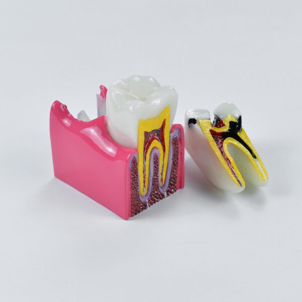
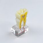
6 Times Caries Model
Original price was: $40.00.$29.88Current price is: $29.88.
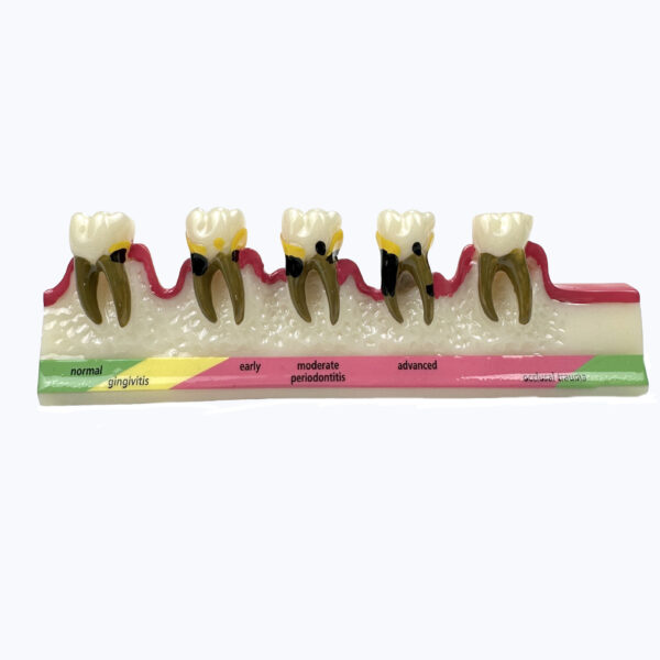
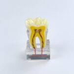
Periodontal Disease Model
Original price was: $50.00.$31.00Current price is: $31.00.
-22%


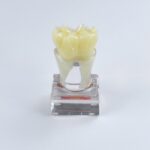


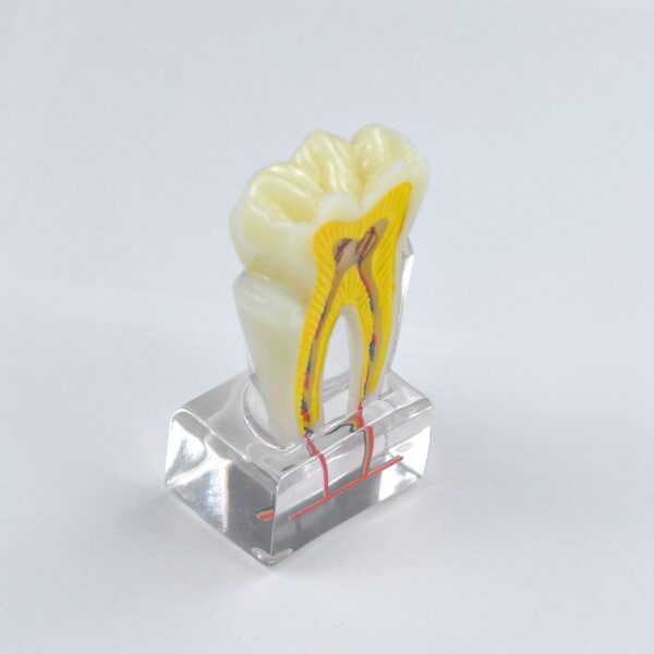
Mixed Dentition Model
Translucent Dental Mixed Dentition Model For Professionals And Students Accurate Analysis.
The Model is an indispensable tool for anyone looking to improve their mixed dentition analysis skills. Its attention to detail, realistic articulation, and durable construction make it an excellent investment for dental students and professionals alike.
Free shipping.


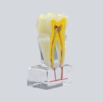
Implant Replacement Module
Dental Implant Replacement Module Model.
Training purpose: drilling training, implant placement, open suture, and gypsum extraction.
Unique design: The model missing tooth with red gingival and bone alternative replacement is a valuable investment for dental practices and students looking to improve their dental implant placement skills.
Free shipping.